Blog

GENDEX GXDP-700 X-RAY SYSTEM - TRACK AND WIN!

Posted by Gendex News on Sep 7, 2012 9:48:00 AM

The month has finally come where we reveal the much anticipated Gendex GXDP-700 Panoramic X-ray sweepstakes winner! Thousands of contestants have been entering to win and the excitement has helped build a fan base for the Experience Gendex Tour that has taken this sweepstakes on the road.

The Experience Gendex North American Tour has housed the 3-in-1 digital system even before the first doctor’s office. As dental professionals around the country stepped aboard the 46ft. “rolling blue thunder” bus, there was no denying the presence of the gorgeous GXDP-700. The sleek style and ergonomic design were instant head turners and when the doctors learned they could have the digital system in their office for the price tag of FREE, everyone jumped at the opportunity. 

 gxex-gxdp-700.jpg

Figure: The GXDP-700 onboard of the Experience Gendex Bus

Tens of thousands of miles were driven to showcase the equipment. Thousands of doctors entered to win the sweepstakes. And now thousands wait for the bus as it sets out on its last leg of the North American tour. The bus will complete its’ final mileage as it rolls into the winning doctor offices parking lot to present them with their GXDP-700 series. If you are one of those anticipating doctors, you can track us live as we travel the country over the next few weeks. The winning stop will be made the last week in September. Perhaps we are closing in on your office!

UnitedStatesMap.png

Knowing the excitement that graced the faces of the thousands who stepped aboard the bus to see and learn about the product, we can only imagine the thrill on the face of the winning contest and their staff as they see the Gendex blue bus pull up to their office. Hold on to your hats, this last month will be an exciting ride.

sensorwinner.jpg

More »

Topics: Digital Technology, GXDP-700 Pan/Ceph/3D unit

NEW AAOMR GUIDELINES FOR 3D IMAGING USE IN IMPLANT PLANNING

Posted by Gendex News on Jun 15, 2012 7:46:00 AM

Over the last few years, Cone Beam Computed Tomography (CBCT), also known as dental 3D imaging, has become more widely adopted in the dental offices. The information delivered by this technology creates invaluable data for the treatment and implant planning process.

Recently, the American Academy of Oral and Maxillofacial Radiology (AAOMR) updated its guidelines for dental imaging in implant treatment, suggesting cone beam CT as the preferred method for presurgical assessment of dental implant sites. Their recommendations are not mandatory; however, their goal is to give dental professionals the qualified opinion on imaging while reducing radiation risks to the patient.

AAOMR advises dentists to use a cone beam CT scanner during any augmentation or implant procedure in order to include cross-sectional imaging orthogonal to the site of interest. CBCT imaging provides an extensive amount of information for the clinician, such as bone structure, bone density, root positioning, fractures, and clear measurements of the anatomical structure. Both doctors and patients alike can now visibly understand the precise diagnosis, method of entry, treatment and recovery.

The AAOMR is continually researching the best methods for dental procedures. The current updated guidelines support the benefits of cross-sectional imaging for implants, which is best achieved with CBCT.

The Gendex GXDP-700 Panoramic 3D imaging system provides the necessary valuable information required for these types of diagnostic procedures while limiting the exposed are to a targeted 4x6cm or 6x8cm volume. This COMBO x-ray system captures both advanced panoramic projections and 3D scans with the touch of a button. The PerfectScout™ system enables 3D scans to zone for the exact area of interest. The GXDP-700™ offers 33 panoramic options — 11 projections for three patient sizes, two 3D volume sizes plus a dose-saving scout view, and the ability to add cephalometrics — 15 options, 5 projections for three patient sizes.


More »

Topics: CBCT cone beam technology, GXDP-700 Pan/Ceph/3D unit

DENTAL PANORAMIC IMAGES AND BONE HEALTH

Posted by Gendex News on Dec 22, 2011 2:27:00 PM

Many factors are involved with keeping teeth in good condition. Besides brushing and flossing, changes in the body chemistry during certain times of life can also affect the dental health. A report on the new study published in the journal Nature Reviews Endocrinology discusses the possibility that dental x-rays can be used to investigate the bone structure in the lower jaw, and predict who is at greater risk of fractures in the future.

The data comes from a reportedly “globally unique” study of women in Gothenburg, Sweden.  Over a span of 43 years, researchers at the University of Gothenburg's Sahlgrenska Academy examined 731 women several times during the study. X-ray images of their jaw bone were analyzed in 1968 and 1980, with special attention to the incidence of subsequent fractures.  The study concluded that the jaw’s bone structure “was sparse in around 20% of the women aged 38-54” at the first examination, and that these women were at significantly greater risk of fractures. The report continues that “The study also shows that the older the person, the stronger the link between sparse bone structure in the jaw and fractures in other parts of the body.”

Maintaining proper nutrition and regular dental visits are proactive ways to detect health issues before they progress. By capturing images of the upper and lower jaw, a panoramic x-ray can discover bone abnormalities, TMD, jaw tumors, cysts or trauma, fractures or impacted teeth, and lead to effective diagnosis and treatment of bone and gum disease, oral cancers, and joint or sinus deformities.  Grethe Jonasson, researcher at the Research Centre of the Public Dental Service in Västra Götaland initiated the fractures study, called “A prospective study of mandibular trabecular bone to predict fracture incidence in women: A low-cost screening tool in the dental clinic.” Jonasson explained, “Dental x-rays contain lots of information on bone structure. By analyzing these images, dentists can identify people who are at greater risk of fractures long before the first fracture occurs.” New pans from Gendex—the  GXDP-700 and GXDP-300 provide practitioners with clear, sharp scans to advance diagnostic capabilities. While many factors affect bone density and the condition of the dentition, panoramic imaging is an effective way for dentists to monitor patients’ quality of life and make constructive recommendations for better dental health well into their senior years.

gxblog4-resized-600.jpg

More »

Topics: Panoramic X-ray Imaging, GXDP-700 Pan/Ceph/3D unit

IN THE PROPER POSITION: DIGITAL PANORAMIC X-RAY UNIT

Posted by Gendex News on Dec 14, 2011 2:24:00 PM

Panoramic x-rays have a long history in dentistry.  In the History of panoramic radiology, author Dr. Dorrit Hallikainen, MD, PhD, notes that “Panoramic radiography is an essential element in oral radiology today.” He continues, “the first attempts to image the whole jaw were made with intraoral radiation sources at the beginning of this century,” and cites that the narrow-beam principle was described as early as 1922, but the equipment was not offered for commercial use until the 1960s.

The pan has evolved into a valuable diagnostic tool, and an article called “Focusing on the Image,” in Dimensions of Dental Hygiene says, “the most important component in producing a diagnostically acceptable panoramic image is the patient positioning.” Author Evelyn M. Thomson, BSDH, MS, further notes, “The dental hygienist who is skilled in understanding panoramic equipment operation and pediatric patient management is more likely to produce radiographic images that result in higher diagnostic yields.”

Kelly Pierce, assistant at the office of Drs. Davila & Velazquez in Greenville, NC, says that the positioning on their new Gendex machine is “super easy for me and the patient.”

slideshow-GXDP-700-600-slides-1.jpg
 

She describes, “We just put them in the machine, move their feet into position, set them up with the laser lines, secure their head, and we have the image in a few seconds.”  Here’s a short video on this process.


She notes that she the system has nearly eliminated the need for retakes. “In the almost three months that we have had our Gendex machine, I have had only one retake,” she says.

We, at Gendex, understand the needs of the dental practice. That’s one reason why both the new GXDP-300™ and GXDP-700™ digital panoramic systems have EasyPosition™. With easy-access controls, a sturdy chin support, ergonomically designed hand-grips for patient comfort, fully adjustable head support, and laser alignment lights to accurately guide patient positioning, dental hygienists and assistants can offer their dentists the high-quality and consistent results of “proper” pans.

More »

Topics: Panoramic X-ray Imaging, GXDP-700 Pan/Ceph/3D unit

THE TREND IS...DIGITAL!

Posted by Gendex News on Oct 26, 2011 2:31:00 PM

When it comes to shopping, everyone has a particular style—some research for months, some make quick decisions, and other wait to see what their peers have done. Dental Products Report’s (DPR) Tech Census 2011 has tracked technology trends in the dental office, with some interesting and positive information regarding the digital office. You can check out the survey here.

The survey cites that 73% of respondents consider themselves as “researchers,” checking out hands-on demos and studies to back up their instincts. It seems that most dentists are planners at heart. The survey notes that “just 7% admitted to being impulse shoppers who see something and want to go for it.” Whatever their decision-making style, the survey points out that technology is definitely foremost on practitioners’ minds.

One of the most sought-after types of technology is digital radiography. Of the 15 categories of technologies included in the survey, digital x-ray system/digital sensor ranked in the top four most commonly owned, by 76 percent of the participant dentists.  These top technologies have steadily increased in popularity in recent years. Some respondents even applauded digital x-ray as the item that has given their practice the biggest boost, with intraoral cameras coming in second.

Dentists who decide on GXS-700™ sensors bring a legacy of sensor research and development to their practice—clear, high resolution images, rounded corners and two sizes for comfort for children and adults, and high speed USB connectivity. Dentists’ digital wish lists also extend to CBCT systems—and the GXCB-500™ and the new GXDP-700™ are two excellent choices for diagnosis and treatment planning of caries, root investigation, orthodontics, implants, surgical procedures, and patient education. Click here to see the newest digital pan/3D/ceph in action!

The survey indicates that there is always room for growth in terms of adding more clinical technology to the practice. It also shows that offices are getting more active with popular outlets such as Facebook, Twitter and YouTube to help promote and grow the practice. We hope you “Like” our Facebook page and check in with our blogs to keep up with all of our exciting developments. Team Gendex is always available to help you understand and implement digital radiography in your practice—helping you to keep up with and exceed digital radiography expectations.

More »

Topics: GXDP-300 Panoramic, Digital Technology, GXDP-700 Pan/Ceph/3D unit

OUR NEW DIGITAL PANORAMIC MACHINES: EASY AS 1-2-3!

Posted by Gendex News on Sep 2, 2011 2:35:00 PM


Easy does it, easy peasy, easy-to-use…for dental imaging products, “easy” means completing a task with the least amount of effort.  And when it comes to touchscreen development, Gendex worked tirelessly on the screens for the GXDP-300 and GXDP-700, the dental digital imaging products. The idea was to make them “intuitive.” I guess you could say that the end goal was to have a screen that takes the least amount of effort to take the image and also saves brain power. 

So, what goes into making these screens intuitive? State-of-the-art electronics and VOC—Voice of the Customer. Not leaving it solely in the hands of knowledgeable engineers, the development team sought design advice from clinicians, the folks that will actually use the systems in the dental practice on a daily basis. 

The team presented various screens to these practitioners, and asked them to demonstrate how they would “take a pan or 3D scan” without giving any hints as to how the screen actually works. In this way, they could tell if the icons and the movement through the different panels really make sense. It’s a process of observing users, making changes, observing users again, making changes again…until the optimal design is achieved. 

As you can see, this VOC process is not quick or easy (no pun intended). It takes time and effort. But in the long-run, Gendex ended up with a graphic user interface for each system, fondly called “GUI” (gooey), that is just another way the company keeps dental teams easily moving through their workdays.

Above is a picture of one of the GUI screens for the GXDP-700, and here’s a link to show the GXDP-300’s GUI in action. Be sure to check out all the videos on the Gendex YouTube Channel!

More »

Topics: GXDP-300 Panoramic, GXDP-700 Pan/Ceph/3D unit

UPGRADING MY GENDEX PANELIPSE PANORAMIC TO CONE BEAM 3D

Posted by Gendex News on Aug 20, 2011 9:55:00 AM

Are you thinking about upgrading your Gendex Paneliplse x-ray machine? You know your Panelispe has served you and your practice well, but you do feel that it is now time to move up and take advantage of the latest technology in dental digital x-ray imaging. If you are contemplating upgrading your Gendex Panelipse panoramic x-ray system, and are somewhat undecided about which way to go, the following reading might stir you in the right direction.
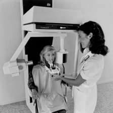
gxpanellipse_ii_w-patient_small.jpegFigure 1: Gendex Panelipse II

Dr. Bryan L. Couch was in a similar spot. His dental office in Coppell, TX is at the forefront of dental digital technology, incorporating the latest in CAD/CAM and digital imaging technology. His trusted Gendex Panelipse panoramic unit was the last piece of the digital puzzle to figure out. The end result of his research led him to replace his Gendex Panelipse with the latest Gendex Pan and Cone Beam marvel: the GXDP-700 S Pan/3D unit. 

dr-couch-and-his-gxdp-700-413x287.jpg

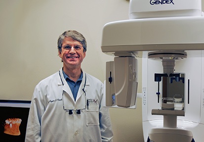
Figure 2: Dr. Couch and his GXDP-700 S Pan/3D unit

We sat down with Dr. Couch, and he was extremely kind to share his story with us. Here is a series of questions we asked: 

Why did you choose the Gendex brand?

All other pieces of equipment in my office were already in digital format. My old panoramic machine, a Gendex Panelipse®, was a trusted workhorse. Although it was still in working condition, I wanted to switch from a traditional film unit to a digital format. I was curious about advancements in imaging technology offered by modern digital panoramic x-ray machines. It offered reduced radiation with flexibility for different projections. The model that I chose had a 3D component, increasing its value to my patients and practice even more.

Why did you choose to add a Gendex GXDP-700 to your practice?

At first, my intention was to use the pan-3D system to help with implant placement.  However, after realizing how advantageous it is to view my patient’s anatomy in three dimensions, I no longer felt comfortable using my older film pans for implant cases because I didn’t feel that I was obtaining the quality diagnostic information necessary for accurate diagnosis and planning.

Implant and Restoration plan.png

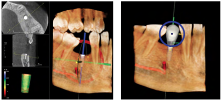
Figure 3: Implant and Restoration plan with GXDP-700 3D scan and InVivo 5 Software

How has your GXDP-700 helped you to expand your practice?

My new pan-3D unit gives me confidence, predictable diagnostics, and results in better treatment. With the 3D option, I can capture a volume of information that I need to confidently place implants, to appropriately and accurately prepare for extractions, to perform root evaluations, solve “mystery endo” cases, and to obtain anatomical documentation for collaboration between me and specialists.

Nerve Mapping.png

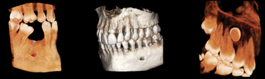
Figure 4: Nerve Mapping, Root Investigations using GXDP-700 Cone Beam scans

What other digital equipment do you already use?

I already have digital intraoral x-ray sensors, digital cameras, and CAD/CAM digital impressions - the E4D™ system.

How does having a digital system improve communication with your referring colleagues?

Consulting doctors are also very grateful to have such detailed information. With traditional film x-ray, the quality is not consistent from office to office. When I electronically send a pan or scan, I know that the specialist and I are both seeing the same quality image. 

Does having a digital system impact patients?

Yes. Patients are so familiar with technology in every aspect of their lives, from phones to computers, and they are used to viewing information on the computer screen.  When I used my film pan, explaining an implant case or other conditions seemed to confuse or even intimidate some patients. Now, I can zoom in on the crisp 2D digital pan image or 3D scans on my large monitor, point out even the smallest details, and in the case of 3D scans, slice and rotate the images to gain the right perspective for education. They appreciate that I have invested in the tools to provide them with individualized treatment.

If the next path forward in upgrading your Panelispe x-ray machine is a feature-rich cone beam unit like the GXDP-700, or simply a rock-solid digital panoramic with TMJ and bitewing capabilities like the GXDP-300 Digital Panoramic, be assured that Gendex offers multiple options in digital panoramic x-ray technology to fit your clinical needs and budget. 

To learn more about the GXDP-700 Pan/Ceph/3D x-ray unit, you can download the product brochure.

Your Imaging Future Starts Today!  
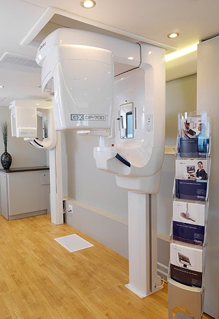
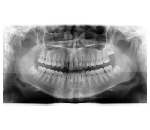
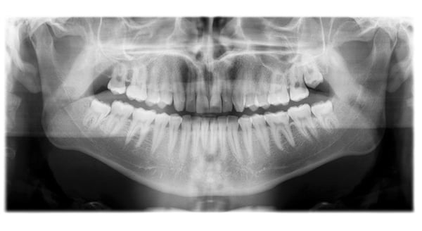
More »

Topics: Digital Technology, CBCT cone beam technology, GXDP-700 Pan/Ceph/3D unit