Blog

KaVo Kerr Announces Dates and Speaker Line-up for Dental 3D University 2017

Posted by Gendex News on Jul 7, 2017 12:48:19 PM

World-class educational event focuses on Cone Beam 3D imaging, how it can enhance your ability to treat patients and expose what might go undetected if limited to 3D imaging.

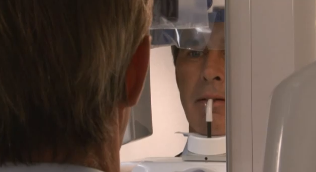
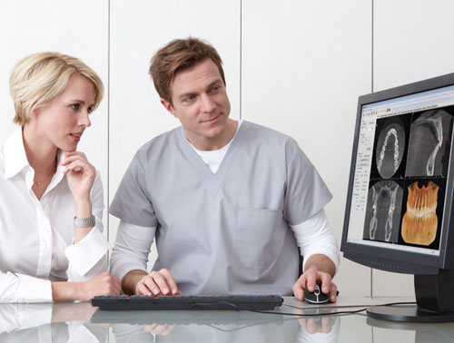
KaVo Kerr is thrilled to announce that it will host its third annual Dental 3D University (3DU), featuring imaging solutions from KaVo™, Instrumentarium™ and Gendex™. 3DU is a one-of-a-kind 2-day event that offers dental professionals an educational environment dedicated to Cone Beam 3D (CBCT) solutions that will enhance their practice and put them in full control of treatment outcomes. This year, 3DU will be held September 29-30 in Dallas, TX.

Welcoming doctors, staff and dental students, 3DU offers attendees the opportunity to earn up to 11 CE Credits. The two-day event will cover topics such as successfully incorporating 3D into your practice and maximizing its investment, implant planning with 3D and surgical guides, reading 3D scans, and more, delivered by world-class speakers and industry experts including: 
  • Dr. Lou Shuman
  • Dr. Kevin Aminzadeh
  • Dr. Douglas Chenin
  • Dr. Lou Graham
  • Dr. Diwakar Kinra
  • Dr. Lisa Koenig
  • Christine Taxin
  • Dr. Terry Work
  • Dr. Gy Yatros
  • Laura Howerton, RDH
  • Art Curley
Taking time away from the practice is a significant investment, but a necessary one in order to acquire the knowledge and education to help dentists maintain a clinical edge. Dentists and their teams will emerge invigorated and energized from the event and ready to leverage the power of 3D technology. KaVo Kerr, bringing together two global leaders under the common banner of Dental Excellence, are in a position to be the premier partner to dentists, providing streamlined services and support for this vital technology, helping dentists and their practices realize their potential. 

“Being the go-to partner for dentists means offering more than just a stellar product. 3DU is an expression of our commitment to the dental team, wanting them to be educated and empowered to use state-of-the-art technology in delivering optimal care and outcomes,” said Pankaj Jaggi, Senior Director, Marketing, for KaVo Kerr Imaging. “We are excited to present a program that will offer tips dentists can put into use right away, as well as concepts that will benefit them in long-term strategizing for the dental practice.”

This impressive line-up deserves a world-class stage, and the venue for 3DU will not disappoint. Located just 20 minutes north of downtown Dallas, the Omni Frisco Hotel is described as “a modern cornerstone of the city reflecting the culture and energy of the area.” Just outside is The Star, the new social hub of Frisco and Home of the Dallas Cowboys, where guests can easily access both entertainment and shopping. Make your hotel arrangements by August 31 to take advantage of the 3DU special rate.

Visit Dental3DU.com today to pre-register for the event and lock-in discounts for both dentists and dental team members.

About KaVo Kerr

KaVo Kerr is a cohesive organization comprised of two global leaders, united to provide dental excellence and serve as a single premier partner for the dental community. KaVo Kerr operates with a common vision inspiring and helping our customers, their patients and our own associates realize their potential. KaVo Kerr offers solutions for endodontics, restoratives, treatment units, infection prevention, imaging, rotary and instruments.

More »

Topics: dental imaging solutions, CBCT cone beam technology, Dental 3D University

GENDEX'S LATEST DIGITAL IMAGING SOLUTIONS AT THE MIDWINTER MEETING

Posted by Gendex News on Feb 23, 2013 6:28:00 PM

Gendex was a must-visit booth this year at the 2013 Chicago Dental Society’s Midwinter Meeting for doctors and staff eager to incorporate the digital imaging technology into their practices.

“It’s been fantastic!” said Peter Thomson, Gendex Territory Sales Manager, about the Midwinter Meeting. “It’s been a great turnout. We’ve had a lot of great questions from doctors; they’re so interested in imaging technology, coming by with their staff to see what’s new from Gendex.” The technology most doctors and staffs were particularly interested in exploring this year can be found in panoramic and cone beam 3D (CBCT) systems like the GXDP-700™ and digital intraoral sensors like the GXS-700™.

Dentists and staffs were particularly interested in digital panorex systems, says Erik Beard, Territory Sales Manager for Gendex. “They look at it as a new solution to expand the range of services and treatments provided to patients," Beard said. "They know it’s so much faster to take a panoramic than a full mouth series, so they are interest to see how digital panoramic images can complement intraoral x-rays.” Throughout the conference, Gendex representatives demonstrated the features of Gendex solutions and explained the ultimate benefits they bring to patients, doctors and staffs.

They’re also ready to answer attendee questions like, “How adding panoramic imaging increase productivity in my practice?” They can even explain how Gendex systems compare to the competition: “Very well,” answers Justin Dickinson, Gendex Territory Sales Manager. “We’ve got award-winning products that exemplify our commitment to continuing innovation and provide dependable performace” he says, explaining that patient comfort and workflow efficienciew are a major focus.

To learn more about the GXDP-700™ pan/ceph/3D and the GXS-700™ digital intraoral sensors, contact Gendex today.


More »

Topics: Panoramic X-ray Imaging, dental imaging solutions, GXDP-700 Pan/Ceph/3D unit, Pan/Ceph/3D, Gendex Team

DIGITAL PANOREX X-RAY MACHINE: HOW TO TAKE GREAT PANORAMIC IMAGES

Posted by Gendex News on Jan 21, 2013 3:05:00 PM

Some people say that position is everything in life — and for capturing quality panoramic images, that is truly the case! Excellent image quality results from careful attention to positioning, and special features on the GXDP-300™ help clinicians to capture panoramics more easily and efficiently.

In just 5 minutes, the video below will show you how to take optimal panoramic x-ray images:

The process starts by setting up the acquisition within your imaging software. Then, ready the unit by positioning the chin rest and bite guide and fully opening the head supports. After pressing the reset button to position the rotating unit, select the projection and patient size on the touchscreen.

Now, for patient positioning. Before settling the patient into place, tell them to remove any glasses, false teeth, jewelry, hair clips, or pins, and then place the protective apron. Press the up and down buttons on the unit to adjust the height of the chin rest so it is slightly higher than the patient’s chin. Ask the patient to step into the unit and grab the handgrips. After the patient places their chin on the chin rest, ask them to position their upper and lower anterior teeth into the groove on the bite guide.


Laser technology is responsible for the next step in positioning. After asking the patient to close their eyes, press the laser button to initiate the laser alignment lights. For mid-saggital placement, verify that the patient’s head is not tilted or turned to one side. For standard panoramic horizontal placement, use the up and down buttons to bring the patient’s Frankfort plane parallel to the laser. For bitewing projection, horizontal placement, use the up and down buttons to bring the patient’s occlusal plane parallel to the laser. Laser lights can be raised or lowered before making adjustments to the Frankfort or occlusal planes. For standard panoramic placement, adjust the chin rest, posteriorly or anteriorly so the cuspids coincide with the laser. For bitewing projection placement, adjust the chin rest to the bitewing marker.

After positioning the laser, carefully push the unit’s head support towards the patient, and rotate the head-support knob clockwise to close the temple supports for a gentle, yet firm grip on the patient’s head. While the patient holds the handgrips firmly, support the patient’s head position by putting your hand on the back of their head, and ask the patient to step forward slightly. Check that the patient is still in the correct position. Ask the patient to press their lips together and to press their tongue against the roof of the mouth.

Positioning for a Panorex

Tip: Looking at a fixed point in the mirror will help the patient to remain still.

During exposure, the unit will rotate around the patient’s head until finished. Exposure should take about 12 seconds for an adult panoramic and 6 seconds for a bitewing. During exposure, move at least seven feet away from the unit, to a place where you are still able to see and hear the patient.

Finally, release the head support, and help the patient out of the unit.

With the GXDP-300, the EasyPosition™ system and instructive alignment guides makes positioning patients of all sizes a smooth and simple process. The touchscreen control panel makes panoramic x-ray capture as easy as 1-2-3:

  1. Select the imaging projection
  2. Choose the patient size
  3. Take the scan

Proprietary FOX™ technology facilitates well-defined, high-quality images with consistent magnification and image uniformity. Panoramic images can provide accurate views of patient anatomy for better diagnostics and a more streamlined workflow and capture process. The GXDP-300 blends performance and simplicity — and puts you in a better position to achieve quality panoramic imaging to provide the best treatment planning for your patients.

For more info on the Gendex GXDP-300, visit the product page

More »

Topics: Panoramic X-ray Imaging, GXDP-300 Panoramic, Digital Technology, dental imaging solutions, Panorex

GENDEX VOTED BEST INTRAORAL X-RAY. 10 YEARS IN A ROW.

Posted by Gendex News on Dec 16, 2012 11:22:00 AM

Since 2003, hundreds of Dentaltown members, also known as Townies, have come together to select dental products worthy enough to be recognized with the prestigious Townie Choice Awards.

Gendex has been a Townie favorite from the very beginning, and the recipient of a Townie Choice Award every single year for 10 years in a row. This year, with 143 votes, nearly 3 times higher than the second classified, Townies have continued in their tradition of assigning the category award for best intraoral x-ray machine to Gendex.

No other dental imaging manufacturer has had the honor of being selected for 10 years in a row! This outstanding series of awards is a testament to the Gendex legacy and its history of dental innovations, which started back in 1893. The company commitment to excellence has resulted in iconic and solid intraoral x-ray products like the popular CDX and GX-770. The current expert DC intraoral x-ray machine continues the Gendex traditions of delivering dental practitioners with proven dependability, clear diagnostic images, and efficient workflow.  

best intraoral dental x-ray


Today, Gendex Dental Systems offers a comprehensive portfolio of dental digital radiography solutions that spans from the award-winning GXS-700 digital x-ray sensors, to extraoral digital panorex machines able to deliver consistent imaging results with clear and easy to acquire panoramic and cephalometric views, as well as dental CBCT scans.  


More »

Topics: dental imaging solutions, Reviews

THE IMPACT OF DENTAL IMAGING TECHNOLOGY

Posted by Gendex News on Jan 27, 2012 2:26:00 PM

Do you remember the first time you saw a big, flatscreen TV? It surely amazed you and all who were lucky enough to see it in action. Technology is engaging, illuminating, and inspiring. And technology in the dental office has the same impact—it can lead to greater understanding, less invasive treatment, and a better overall perception of the office.

In this economy and competitive atmosphere, technology can also set you apart. Many dental educators agree that technology builds patient loyalty, and this loyalty is a large part of the success of dentists as practitioners and business owners.

So what are patients looking for? A recently published article coached patients on questions to ask their potential new dentist. Rated right alongside, “Where did you receive your dental training?” patients also were advised to ask:

  1. “What is the most recent technology that you have added to your practice?”
  2. “If your fees are more expensive, what makes your practice a good investment?”
  3. “What services do you provide in your area that other dentists do not?”
  4. Digital Imaging can provide a positive answer to all three of these questions. 

On a recent DPR Friday weekly video broadcasts, Director of Content for Advanstar Dental Media, Thais Carter, poses the question, “Have you ever wondered what patients think about the technology in your office?”  This video gives a bit of patient Amanda Davis’ perspective on her recent visit to the office of one of our Gendex owners, Dr. Brian Couch in Coppell, Texas.

The full article, “The Multi-Layered Comfort of 3D Technology,” yields more insights into Amanda’s experiences with the new Gendex digital pan-3D system and with Dr. Couch.  

Find out more about our technology at www.gendex.com.

impactofdentalimaging-resized-600.jpg

More »

Topics: dental imaging solutions

THE ROOT OF THE MATTER: 3D DENTAL IMAGING

Posted by Gendex News on Dec 7, 2011 2:29:00 PM

Increasingly, dentists are turning to 3D imaging to find conditions that would be undetectable on 2D radiographs.  A joint position statement by the American Association of Endodontists (AAE) and the American Association of Oral and Maxillofacial Radiography (AAOMR) noted that “Significantly increased use of CBCT is evidenced by a recent Web-based survey of active AAE members in the United States and Canada, which found that 34.2% of 3,844 respondents indicated that they were utilizing CBCT.” Most frequently, the 3D option was chosen for the “diagnosis of pathosis, preparation for endodontic treatment or endodontic surgery, and for assistance in the diagnosis of trauma-related injuries.”

A recent article in Dental Traumatology noted that 3D is a beneficial tool for diagnosing vertical root fractures (VRFs). Since the diagnosis is based on clinical and radiographic presentations, the study notes, “It might be difficult to detect VRFs in non-endodontically treated molars by conventional radiographs in certain situations because of the limitations of 2D images and many others factors such as the VRFs being in their early stages.”  Variations in root shapes and the position of the VRF can affect detection on conventional radiographic methods, and in turn, may lead to a misinterpretation of a radiolucent fracture line. The study presents four case reports to illustrate how CBCT was utilized to gather more information of the presence of VRFs, and “successfully diagnose VRFs based on direct visualization of radiolucent lines, especially those suspected from routine conventional radiographs, as well as their symptoms and clinical findings.”

To get to the root of the matter, or at least the fracture on the root, CBCT scans provide high definition diagnostic images for efficient treatment planning.


More »

Topics: dental imaging solutions, CBCT cone beam technology