Some people say that position is everything in life — and for capturing quality panoramic images, that is truly the case! Excellent image quality results from careful attention to positioning, and special features on the GXDP-300™ help clinicians to capture panoramics more easily and efficiently.
In just 5 minutes, the video below will show you how to take optimal panoramic x-ray images:
The process starts by setting up the acquisition within your imaging software. Then, ready the unit by positioning the chin rest and bite guide and fully opening the head supports. After pressing the reset button to position the rotating unit, select the projection and patient size on the touchscreen.
Now, for patient positioning. Before settling the patient into place, tell them to remove any glasses, false teeth, jewelry, hair clips, or pins, and then place the protective apron. Press the up and down buttons on the unit to adjust the height of the chin rest so it is slightly higher than the patient’s chin. Ask the patient to step into the unit and grab the handgrips. After the patient places their chin on the chin rest, ask them to position their upper and lower anterior teeth into the groove on the bite guide.
Laser technology is responsible for the next step in positioning. After asking the patient to close their eyes, press the laser button to initiate the laser alignment lights. For mid-saggital placement, verify that the patient’s head is not tilted or turned to one side. For standard panoramic horizontal placement, use the up and down buttons to bring the patient’s Frankfort plane parallel to the laser. For bitewing projection, horizontal placement, use the up and down buttons to bring the patient’s occlusal plane parallel to the laser. Laser lights can be raised or lowered before making adjustments to the Frankfort or occlusal planes. For standard panoramic placement, adjust the chin rest, posteriorly or anteriorly so the cuspids coincide with the laser. For bitewing projection placement, adjust the chin rest to the bitewing marker.
After positioning the laser, carefully push the unit’s head support towards the patient, and rotate the head-support knob clockwise to close the temple supports for a gentle, yet firm grip on the patient’s head. While the patient holds the handgrips firmly, support the patient’s head position by putting your hand on the back of their head, and ask the patient to step forward slightly. Check that the patient is still in the correct position. Ask the patient to press their lips together and to press their tongue against the roof of the mouth.
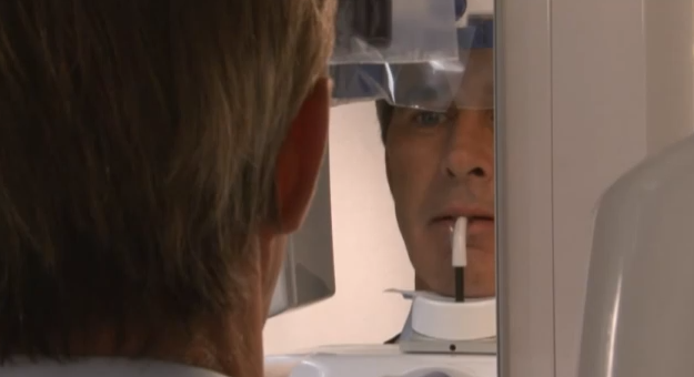
Tip: Looking at a fixed point in the mirror will help the patient to remain still.
During exposure, the unit will rotate around the patient’s head until finished. Exposure should take about 12 seconds for an adult panoramic and 6 seconds for a bitewing. During exposure, move at least seven feet away from the unit, to a place where you are still able to see and hear the patient.
Finally, release the head support, and help the patient out of the unit.
With the GXDP-300, the EasyPosition™ system and instructive alignment guides makes positioning patients of all sizes a smooth and simple process. The touchscreen control panel makes panoramic x-ray capture as easy as 1-2-3:
- Select the imaging projection
- Choose the patient size
- Take the scan
Proprietary FOX™ technology facilitates well-defined, high-quality images with consistent magnification and image uniformity. Panoramic images can provide accurate views of patient anatomy for better diagnostics and a more streamlined workflow and capture process. The GXDP-300 blends performance and simplicity — and puts you in a better position to achieve quality panoramic imaging to provide the best treatment planning for your patients.
For more info on the Gendex GXDP-300, visit the product page.
